Читайте также:
|
SCLERA
The fibrous, external layer of the eyeball protects the more delicate internal structures and provides sites for muscle insertion. (In reference to the eye, the terms "external/outer" and "internal/ inner" refer to structures closer to the eyeball's surface or in its interior, respectively.) The opaque white posterior five-sixths of the external layer is the sclera; this forms a segment of a sphere with a diameter of approximately 22 mm in adults. The sclera averages 0.5 mm in thickness, is relatively avascular, and consists of tough, dense connective tissue containing flat type I collagen bundles which intersect in various directions while remaining parallel to the surface of the organ, with a moderate amount of ground substance and scattered fibroblasts. Tendons of the extraocular muscles that move the eyes insert into anterior areas of the sclera. Posteriorly the sclera thickens to approximately 1 mm and joins with the epineurium covering the optic nerve. A thin inner region of the sclera, adjacent to the choroid, is slightly less dense, with thinner collagen fibers, more fibroblasts, elastic fibers, and melanocytes.
CORNEA
In contrast to the sclera, the anterior one-sixth of the eye—the cornea —is colorless, transparent, and completely avascular. A section of the cornea shows that it consists of five layers:
- an external stratified squamous epithelium,
- an anterior limiting membrane (Bowman's membrane, the basement membrane of the stratified epithelium),
- the stroma,
- a posterior limiting membrane (Descemet's membrane, the basement membrane of the endothelium), and
- an inner simple squamous endothelium..
The stratified surface epithelium is nonkeratinized, with five or six cell layers of cells comprising about 10% of the corneal thickness. Numerous mitotic figures are present in the basal layers, particularly near the periphery of the cornea, reflecting the epithelium's high capacity for cell renewal and repair. The flattened surface cells have microvilli and folds protruding into a protective layer or tear film of lipid, glycoprotein, and water about 7 m thick. As another
protective adaptation, the corneal epithelium also has one of the richest sensory nerve supplies of any tissue. The basement membrane of this epithelium is very thick (8–12 m) and contributes to the stability and strength of the cornea, helping to protect against infection of the underlying stroma.
| CORNEA | |
| Outermost layer (non-keratinized stratified squamous epithelium) | The cells in the deepest layer of the epithelium are columnar; in the middle layers they are polygonal; and in the superficial layers they are flattened. The cells are arranged with great regularity. |
| Anterior limiting lamina | It is made up of collagen fibrils embedded in matrix. |
| Substantia propria (corneal stroma) | It is made up of collagen fibres embedded in a ground substance con- taining sulphated glycosaminoglycans. |
| Posterior limiting lamina | It is a true basement membrane. |
| Endothelium of the anterior chamber | It is a single layer of flattened cells. |


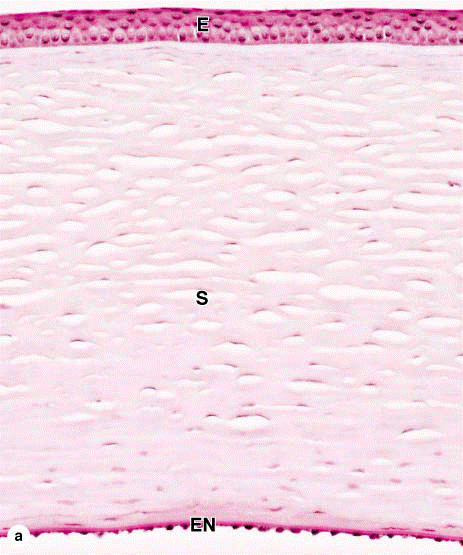
Cornea.
The anterior structure of the eye, the cornea has five layers. (a): The micrograph shows the external stratified squamous epithelium (E), which is nonkeratinized and five or six cells thick. It is densely supplied with sensory free nerve endings that trigger the blinking reflex and its surface is covered with a tear film produced by glands in the eyelids and superior orbit. The stroma (S) comprises approximately 90% of the cornea's thickness, consisting of some 60 layers of long type I collagen fibers arranged in a precise orthogonal array and alternating with flattened cells called keratocytes. The stroma is lined internally by endothelium (EN). X100. H&E.
(b): The corneal epithelium rests firmly on the thick homogeneous Bowman's membrane (arrow). The stroma is completely avascular and nutrients reach the keratocytes and epithelial cells by diffusion from the surrounding limbus and aqueous humor behind the cornea. X400. H&E. (c): The posterior surface of the cornea is covered by simple squamous epithelium (endothelium) that rests on another thick, strong layer of collagen and other extracellular material called Descemet's membrane (arrow). Na/K ATPase of the endothelial cells is responsible for pumping Na+ and drawing water out of the cornea, maintaining its proper state of hydration. In this state the cornea is perfectly transparent and with its curvature is a major refractive structure of the eye. X400. H&E.
The thick stroma, or substantia propria, is formed of approximately 60 layers of parallel collagen bundles that align at approximately right angles to each other
and may cross the complete corneal diameter. The uniform orthogonal array of collagen fibrils contributes to the transparency of this avascular tissue. Between the collagen lamellae are cytoplasmic extensions of flattened fibroblast-like cells called keratocytes. The ground substance surrounding these cells is rich in proteoglycans such as lumican, containing keratan sulfate and chondroitin sulfate, which help maintain the precise organization and spacing of the
collagen fibrils.
The posterior surface of the stroma is bounded by another thick (~10 m) structure (Descemet's membrane) composed of fine, interwoven collagen fibers, upon which lies the corneal endothelium. Cells of this simple squamous epithelium are active in protein synthesis to maintain this basement membrane and in pumping sodium ions into the adjacent anterior chamber. Chloride ions and water follow passively from the corneal stroma. In this way, the endothelium is largely responsible for maintaining a state of hydration within the cornea that helps provide maximum transparency and optimal light refraction.
CHOROID
The choroid is a highly vascular tunic in the posterior two-thirds of the eye, with loose, well-vascularized connective tissue rich in collagen and elastic fibers, fibroblasts, melanocytes, macrophages, lymphocytes, mast cells, and plasma cells. The abundant melanocytes give the layer its characteristic black color and block light from entering the eye except through the pupil.

Дата добавления: 2015-10-30; просмотров: 122 | Нарушение авторских прав
| <== предыдущая страница | | | следующая страница ==> |
| Layers of the eye. | | | Sclera, choroid, and retina. |