Section through a thin portion of the wall of a long-bone diaphysis showing both periosteum (P) and endosteum (E). The periosteum covers bone and provides a supply of osteoprogenitor cells which become osteoblasts for new bone formation. These cells are located in the inner, more cellular layers of the periosteum, next to the bone matrix. Externally the periosteum consists of a thick layer of more fibrous, dense connective tissue, which merges with ligaments and other connective tissues. Perforating fibers fastening the periosteum to the bone matrix are not seen in routine light microscope preparations. The periosteum has a good blood supply, but the cavities lined by endosteum, the marrow cavities, are very rich in blood sinuses and blood-forming tissue. X100. H&E.
The large internal marrow cavities of bone are lined by endosteum. Endosteum is a single very thin layer of connective tissue, containing flattened osteoprogenitor cells and osteoblasts, which covers the small spicules or trabeculae of bone that project into these cavities. The endosteum is therefore considerably thinner than the periosteum.
The principal functions of periosteum and endosteum are nutrition of osseous tissue and provision of a continuous supply of new osteoblasts for repair or growth of bone.
An osteon
An osteon.
In preparations of dried, ground bone osteons can be seen with lacunae (L) situated between concentric lamellae and interconnected by fine canaliculi (C). Although it is not apparent by light microscopy, each lamella consists of multiple parallel arrays of collagen fibers. In adjacent lamellae, the collagen fibers are oriented in different directions. The presence of large numbers of lamellae with differing fiber orientations provides the bone with great strength, despite its light weight. Only remnants of the osteocytes (O) in some lacunae and of the osteonic canal's contents are seen in ground bone. In living tissue osteocytic processes connected via gap junctions are present in successive canaliculi, making cells in all the lamellae in communication with the blood vessels in the central canal. X500.
In each lamella, type I collagen fibers are aligned in parallel and follow a helical course. The pitch of the helix is, however, different for different lamellae, so that at any given point, fibers from adjacent lamellae intersect at approximately right angles. The specific organization of collagen fibers in successive lamellae of each osteon is highly important for the great strength of secondary bone.
In compact bone (eg, the diaphysis of long bones) besides forming osteons, the lamellae also exhibit a typical organization consisting of multiple external circumferential lamellae and often some inner circumferential lamellae. Inner circumferential lamellae are located around the marrow cavity andexternal circumferential lamellae are located immediately beneath the periosteum.
Each osteon is a long, often bifurcated cylinder generally parallel to the long axis of the diaphysis. It consists of a central canal surrounded by 4–10 concentric lamellae. Each endosteum-lined canal contains blood vessels, nerves, and loose connective tissue. The central canals communicate with the marrow cavity and the periosteum and with one another through transverse or oblique perforating canals (formerly known as Volkmann canals). The transverse
canals do not have concentric lamellae; instead, they perforate the lamellae. All osteonic and perforating canals in bone tissue come into existence when matrix is laid down around preexisting blood vessels.
Lamellar bone: Perforating canals and interstitial lamellae.


Lamellar bone: Perforating canals and interstitial lamellae.
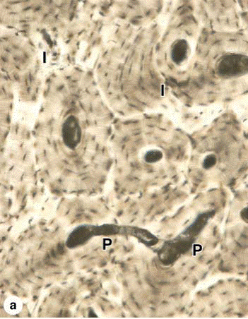
(a): Transverse perforating canals (P) connecting adjacent osteons are shown at the left side of the micrograph. Such canals "perforate" lamellae and provide another source of microvasculature for the central canals of osteons. Among the intact osteons are also found remnants of eroded osteons, seen as irregular interstitial or intermediate lamellae (I). X100. (b): Schematic diagram shows remodeling of compact lamellar bone showing three generations of osteonic haversian systems and their successive contributions to the formation of interstitial lamellae. Remodeling is a continuous process that involves the coordinated activity of osteoblasts and osteoclasts,
and is responsible for adaptation of bone to changes in stress, especially during the body's growth.
Among the osteons between the two circumferential systems are numerous irregularly shaped groups of parallel lamellae called interstitial lamellae. These structures are lamellae remaining from osteons partially destroyed by osteoclasts during growth and remodeling of bone.
BLOOD
BLOOD:
- PLASMA
- CELLULAR (FORMED) ELEMENTS
| PLASMA | A homogeneous, slightly alkaline fluid which contains dissolved gases, inorganic salts, proteins, carbohydrates, lipids, and certain other organic substances. It constitutes fifty-five per cent of blood. |
| CELLULAR (FORMED) ELEMENTS | |
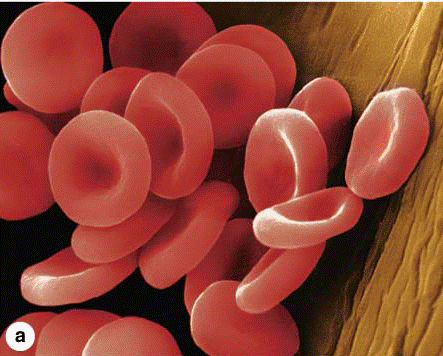
| Erythrocyters | Red blood corpuscles that have lost their nuclei and their cytoplasmic organelles. They contain a red coloured protein called haemoglobin. |
| Leucocytes | |
| - Granulocytes | They have granules in their cytoplasm. They contain many-lobed nucleus. |
| 1. basophils | Their large cytoplasmic granules stain with basic dyes. |
| 2. eosinophils | Their large cytoplasmic granules stain brightly with acid dyes. |
| 3. neurtophils | Their very fine cytoplasmic granules stain lightly with both acidic and basic dyes. |
| - Agranulocytes | They have homogeneous cytoplasm and spherical or reniform nuclei. |
| 1. lymphocytes | They have a relatively large nucleus surrounded by a narrow rim of cytoplasm. |
| 2. monocytes | Large cell containing eccentrically placed nucleus and relatively abundant cytoplasm. |
| Blood platelets | Small protoplasmic discs devoid of nuclei. |


Дата добавления: 2015-10-30; просмотров: 166 | Нарушение авторских прав
| <== предыдущая страница | | | следующая страница ==> |
| Osteoclasts and their activity. | | | Neutrophil ultrastructure. |