|
Читайте также: |
Characteristic features of macrophages seen in this TEM of one such cell are the prominent nucleus (N) and the nucleolus (Nu) and the numerous secondary lysosomes (L). The arrows indicate phagocytic vacuoles near the protrusions and indentations the cell surface. X10,000.
Mast Cells
Mast cells are large, oval or round connective tissue cells, 20–30 m in diameter, whose cytoplasm is filled with basophilic secretory granules. The rather small, spherical nucleus is centrally situated and may be obscured by the cytoplasmic granules.

MAST CELL

Mast cells.
Mast cells are components of loose connective tissues, often located near small blood vessels (BV). (a): They are typically oval-shaped, with cytoplasm filled with strongly basophilic granules. X400. PT. (b): Ultrastructurally mast cells show little else around the nucleus (N) besides these cytoplasmic granules (G), except for occasional mitochondria (M). The granule staining in the TEM is heterogeneous and variable in mast cells from different tissues; at higher magnifications some granules may show a characteristic scroll-like substructure (inset) that contains preformed mediators such as histamine and proteoglycans. The ECM near this mast
cell includes elastic fibers (E) and bundles of collagen fibers (C).
PLASMA CELL

Plasma cells.
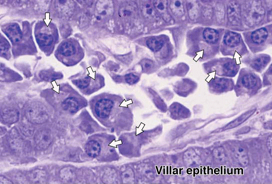
Plasma cells are abundant in this portion of an inflamed intestinal villus. The plasma cells are characterized by their abundant basophilic cytoplasm involved in the synthesis of antibodies. A large pale Golgi apparatus (arrows) near each nucleus is the site of the terminal glycosylation of the antibodies (glycoproteins). Plasma cells can leave their sites of origin in lymphoid tissues, move to connective tissue, and produce the antibodies that mediate immunity. X400 PT.
COLLAGEN FIBRES


Дата добавления: 2015-10-30; просмотров: 129 | Нарушение авторских прав
| <== предыдущая страница | | | следующая страница ==> |
| Functions of connective tissue cells. | | | Dense regular connective tissue. |