
|
Читайте также: |
TEM of a sectioned human neutrophil immunostained for peroxidase reveals the two types of cytoplasmic granules: the small, pale, peroxidase-negative specific granules and the larger, dense, peroxidase-positive azurophilic granules. Specific granules undergo exocytosis during and after diapedesis, releasing many factors with variousactivities, including enzymes to digest ECM components and bacteriostatic factors. Azurophilic granules are modified lysosomes with components to kill engulfed bacteria.
The nucleus is lobulated and the central Golgi apparatus is small. Rough ER and mitochondria are not abundant, because this cell utilizes glycolysis and is in the terminal stage of its differentiation. X27,000.
EOSINOPHILS
Eosinophils are far less numerous than neutrophils, constituting only 2–4% of leukocytes in normal blood. In blood smears, this cell is about the same size as a neutrophil, but with a characteristic bilobed nucleus. The main identifying characteristic is the abundance of large, red specific granules (about 200 per cell) that are stained by eosin.


Eosinophils.
Eosinophils are about the same size as neutrophils but have bilobed nuclei and abundant coarse cytoplasmic granules. The cytoplasm is often filled with brightly eosinophilic specific granules, but also includes some azurophilic granules. (a): Micrograph shows an eosinophil next to a neutrophil for comparison with its nucleus and granules. X1500. Wright. (b): Even with granules filling the cytoplasm, the two nuclear lobes of eosinophils are usually clear. X1500. Giemsa. (c): TEM of a sectioned eosinophil clearly shows the unique specific granules, as oval structures with disk-shaped electron-dense crystalline cores (EG). These along with lysosomes and a few
mitochondria (M) fill the cytoplasm around the bilobed nucleus (N). X20,000.
BASOPHILS
Basophils are also about 12–15 m in diameter, but make up less than 1% of blood leukocytes and are therefore difficult to find in smears of normal blood. The nucleus is divided into two or more irregular lobes, but the large specific granules overlying the nucleus usually obscure its shape.
The azurophilic specific granules (0.5 m in diameter) stain dark blue or metachromatically with the basic dye of blood smear stains and are fewer and more irregular in size and shape than the granules of the other granulocytes. The metachromasia is due to the presence of heparin and other
sulfated glycosaminoglycans (GAGs) in the granules. Basophilic specific granules also contain much histamine and various mediators of inflammation, including platelet activating factor, eosinophil chemotactic factor, and phospholipase A which produces low molecular weight factors called leukotrienes.


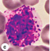

Basophils.
(a, b, c): Basophils are approximately the same size as neutrophils and eosinophils, but have large, strongly basophilic specific granules which usually obstruct the appearance of the nucleus having two or three irregular lobes. a and b: X1500, Wright; c: X1500, Giemsa. (d): TEM of a sectioned basophil reveals the lobulated nucleus (N), appearing as three separated portions, the large specific basophilic granules (B), mitochondria (M), and Golgi complex (G). Basophils exert many activities modulating the immune response and inflammation and share many functions with mast cells, which are normal, longer term residents of connective tissue. X16,000.
LYMPHOCYTES
Lymphocytes constitute a family of leukocytes with spherical nuclei. They can be subdivided into functional groups according to distinctive surface molecules (markers) that can best be distinguished immunocytochemically, notably T lymphocytes, B lymphocytes, and natural killer (NK) cells.
Lymphocytes have diverse functional roles related to immune defense against invading microorganisms, foreign or abnormal antigens, and cancer cells.
Most lymphocytes in the blood are small with diameters of 6–8 m; medium and large lymphocytes range in size from 9 to 18 m in diameter. Some larger lymphocytes may be cells that have been activated by specific antigens. The small lymphocytes that predominate in the blood are characterized by spherical nuclei, sometimes indented, and condensed, very basophilic chromatin, making them easily distinguishable from granulocytes.
The cytoplasm of the small lymphocyte is scanty, and in blood smears it appears as only a thin rim around the nucleus. It is slightly basophilic and may contain a few azurophilic granules, along with a few mitochondria and a small Golgi apparatus; it contains free polyribosomes.

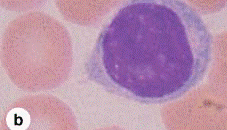


Lymphocytes.
Lymphocytes are agranulocytes and lack the specific granules characteristic of granulocytes. Lymphocytes circulating in blood range in size from 6 to 15 m in diameter and are sometimes classified arbitrarily as small, medium, and large. (a): The most numerous small lymphocytes shown here are slightly larger than the neighboring erythrocytes and often have only a thin rim of cytoplasm surrounding the spherical nucleus. X1500. Giemsa. (b): Medium lymphocytes are distinctly larger than erythrocytes. X1500. Wright. (c): Large lymphocytes, much larger than erythrocytes, may represent activated cells that have returned to the circulation. X1500. Giemsa.
(d): Ultrastructurally a medium-sized lymphocytes is seen to be mostly filled with a euchromatic nucleus (N), with a nucleolus (Nu), surrounded by cytoplasm containing mitochondria (M), free polysomes, and a few lysosomes (azurophilic granules). X22,000.
Lymphocytes vary in life span according to their specific functions; some live only a few days and others survive in the circulating blood or other tissues for many years. They are the only type of leukocytes that, following diapedesis, can return from the tissues back to the blood.
MONOCYTES
Monocytes are bone marrow–derived agranulocytes with diameters varying from 12 to 20 m. The nucleus is large, off-center, and may be oval, kidney-shaped, or distinctly U-shaped. The chromatin is less condensed than in lymphocytes and stains lighter than that of large lymphocytes


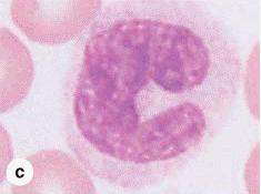
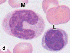
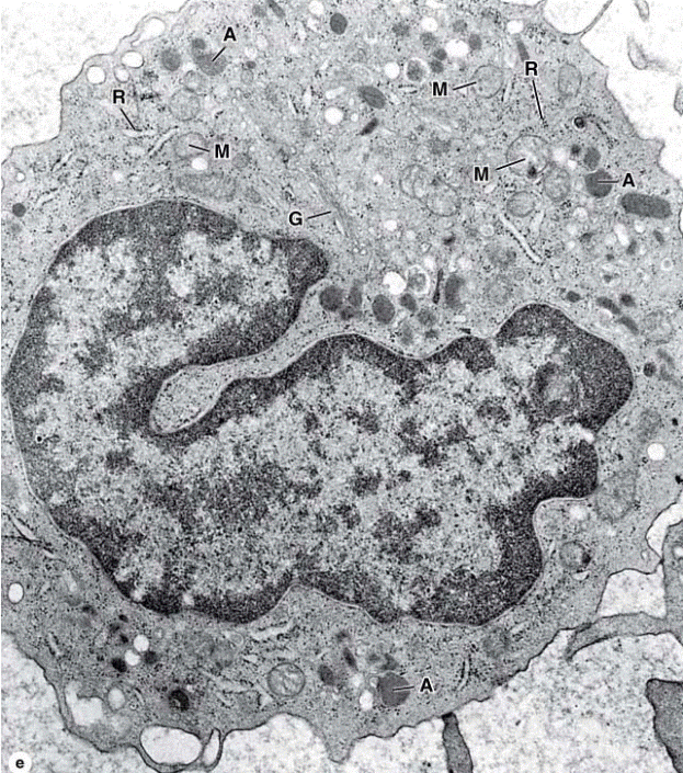
Monocytes.
Monocytes are large agranulocytes with diameters from 12 to 20 m that circulate as precursors to macrophages and other cells of the mononuclear phagocyte system. (a, b, c, d): Micrographs of monocytes that show their eccentric nuclei indented, kidney-shaped, or U-shaped. a: X1500, Giemsa; b–d: X1500, Wright. (e): TEM of thecytoplasm of a monocyte shows a Golgi apparatus (G), mitochondria (M), and lysosomes or azurophilic granules (A). Rough ER is poorly developed and there are some freeribosomes (R). X22,000..
PLATELETS
PLATELETS
Blood platelets (thrombocytes) are nonnucleated, disklike cell fragments 2–4 m in diameter. Platelets originate by fragmentation at the ends of cytoplasmic processes extending from giant polyploid cells called megakaryocytes in the bone marrow. Platelets promote blood clotting and help repair minor tears or leaks in the walls of blood vessels, preventing loss of blood. Normal platelet counts range from 200,000 to 400,000 per microliter of blood. Platelets have a life span
of about 10 days.
In stained blood smears, platelets often appear in clumps. Each platelet has a lightly stained peripheral zone, the hyalomere, and a central zone containing darkerstaining granules, called the granulomere.


Platelets.
Platelets are cell fragments 2–4 m in diameter derived from megakaryocytes of bone marrow. Their primary function is to rapidly release the content of their granules upon contact with collagen (or other materials outside of the endothelium) to begin the process of clot formation and reduce blood loss from the vasculature. (a): In a blood smear, platelets (arrows) are often found as aggregates. Individually they show a lightly stained hyalomere region surrounding a more darkly stained central granulomere containing membrane-enclosed granules. X1500. Wright. (b): Ultrastructurally a platelet typically shows a system of microtubules and actin filaments near the periphery to help maintain its shape and an open canalicular system of vesicles continuous with the plasmalemma. The central granulomere region contains glycogen and secretory granules of different types. X40,000. (c): TEM section shows platelets adhering to collagen (C). Upon adhesion to collagen, platelets exocytose their granules into the
canalicular system, which allows the very rapid secretion of factors involved in blood coagulation. Degranulating platelets (arrows) remain as an aggregate until their contents are exhausted. Other proteins involved in coagulation come from the plasma and from processes of adjacent endothelial cells (EP). The electron-dense structure on the right is part of an erythrocyte. X;7500.
STEM CELLS, GROWTH FACTORS, & DIFFERENTIATION
Stem cells are pluripotent cells capable of asymmetric division and self-renewal. Some of their daughter cells form specific, irreversibly differentiated cell types, and other daughter cells remain stem cells. A constant number of pluripotent stem cells is maintained in a pool and cells recruited for differentiation are replaced with daughter cells from the pool.
Hemopoietic stem cells can be isolated by using fluorescence-labeled antibodies to mark specific cell-surface antigens and a fluorescence-activated cell-sorting (FACS) instrument. Stem cells are studied using experimental techniques that permit analysis of hemopoiesis in vivo and i n vitro.
In vivo techniques include injecting the bone marrow of normal donor mice into lethally irradiated mice whose hematopoietic cells have been destroyed. In these animals, the transplanted bone marrow cells develop colonies of hematopoietic cells in the marrow cavities and spleen. This work led to the development of bone marrow transplants now used clinically to treat potentially lethal hemopoietic disorders.
In vitro techniques involve the use of semisolid tissue culture media made with a layer of cells derived from bone marrow stroma or purified protein growth factors produced by marrow stromal cells. Such methods create microenvironmental conditions sufficiently similar to the normal in vivo conditions to favor hemopoietic stem
cell growth and differentiation.
Дата добавления: 2015-10-30; просмотров: 177 | Нарушение авторских прав
| <== предыдущая страница | | | следующая страница ==> |
| Periosteum and endosteum. | | | The three types of muscle. |