Читайте также:
|
Longitudinal sections reveal the striations characteristic of skeletal muscle. (a): Parts of three muscle fibers separated by very small amounts of endomysium. One fibroblast nucleus (F) is shown. Muscle nuclei (N) are found against the sarcolemma. Along each fiber thousands of dark-staining A bands alternate with l i ghter I bands. X200. H&E. (b): At higher magnification, each fiber can be seen to have three or four myofibrils, with their striations slightly out-of-alignment with one another.
Myofibrils are cylindrical bundles of thick and thin myofilaments which fill most of each muscle fiber. The middle of each I band can be seen to have a darker Z line (or disk). X500. Giemsa. (c): TEM showing the more electron-dense A bands bisected by a narrow, less electron-dense region called the H zone and in the I bands the presence of sarcoplasm with mitochondria (M), glycogen granules, and small cisternae of SER around the Z line. X24,000.


Structure of a myofibril: a series of sarcomeres.
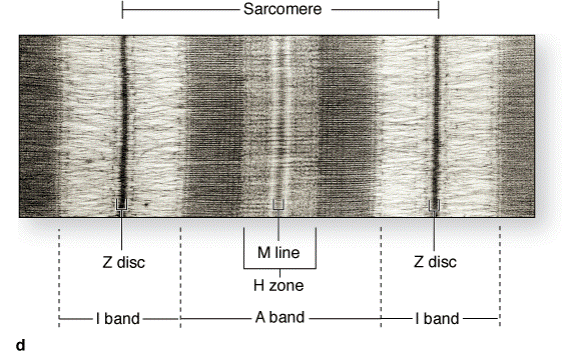
(a): Diagram indicates that each muscle fiber contains several parallel bundles called myofibrils. (b): Each myofibril consists of a long series of sarcomeres which contain thick and thin filaments and are separated from one another by Z discs. (c): Thin filaments are actin filaments with one end bound to -actinin, the major protein of the Z disc. Thick filaments are bundles of myosin, which span the entire A band and are bound to proteins of the M line and to the Z disc across the I bands by a very large protein called titin, which has spring-like domains. (d): The molecular organization of the sarcomeres has bands of greater and lesser protein density,
resulting in staining differences that produce the dark and light-staining bands seen by light microscopy and TEM. (e): TEM cross-sections through different regions of the sarcomere, as shown here, were useful in determining the relationships between thin and thick myofilaments and other proteins, as shown in part b of this figure.
Thin and thick filaments are arranged so that each myosin bundle contacts six actin filaments.
The sarcoplasm has little RER or free ribosomes and is filled primarily with long cylindrical filamentous bundles called myofibrils running parallel to the long axis of the fiber. The myofibrils have a diameter of 1–2 m and consist of an end-to-end repetitive arrangement of sarcomeres. The lateral registration ofsarcomeres in adjacent myofibrils causes the entire muscle fiber to exhibit a characteristic pattern of transverse striations.
The A and I banding pattern in sarcomeres is due mainly to the regular arrangement of two types of myofilaments —thick and thin—that lie parallel to the long axis of the myofibrils in a symmetric pattern.
The thick filaments are 1.6 m long and 15 nm wide; they occupy the A band, the central portion of the sarcomere. The thin filaments run between and parallel to the thick filaments and have one end attached to the Z line. Thin filaments are 1.0 m long and 8 nm wide. As a result of this arrangement, the I bands consist of the portions of the thin filaments that do not overlap the thick filaments (which is why they are lighter staining). The A bands are composed mainly of thick filaments in addition to overlapping portions of thin filaments. Close observation of the A band shows the presence of a lighter zone in its center, the H zone, that corresponds to a region consisting only of the rod-like portions of the myosin molecule with no thin filaments present. Bisecting the H zone is the M line (Ger. Mitte, middle), a region where lateral connections are made between adjacent thick filaments. Major proteins present in the M line region are myomesin, a myosin-binding protein which holds the thick filaments in place, and creatine kinase, which catalyzes the transfer of phosphate groups from phosphocreatine (a storage form of high-energy phosphate groups) to adenosine diphosphate (ADP), thus helping to supply adenosine triphosphate (ATP) for muscle contraction.
Thin and thick filaments overlap for some distance within the A band. As a consequence, a cross section in the region of filament overlap shows each thick filament surrounded by six thin filaments in the form of a hexagon.
Thin filaments are composed of F-actin, associated with tropomyosin, which also forms a long fine polymer, and troponin, a globular complex of three subunits.
Thick filaments consist primarily of myosin. Myosin and actin together represent 55% of the total protein of striated muscle.
F-actin consists of long filamentous polymers containing two strands of globular (G-actin) monomers, 5.6 nm in diameter, twisted around each other in a double helical formation. G-actin molecules are asymmetric and polymerize to produce a filament with polarity. Each G-actin monomer contains a binding site for myosin. Actin filaments, which are anchored perpendicularly on the Z line by the actin-binding protein -actinin, exhibit opposite polarity on each side of the line.
Дата добавления: 2015-10-30; просмотров: 171 | Нарушение авторских прав
| <== предыдущая страница | | | следующая страница ==> |
| The three types of muscle. | | | Structures of neuron. |