Читайте также:
|
The osteoclast is a large cell with several nuclei derived by the fusion in bone of several blood-derived monocytes. (a): Microscopic section showing two osteoclasts (arrows) digesting or resorbing bone matrix in resorption bays on the matrix surface. X400. H&E. (b): Diagram showing each osteoclast has a circumferential zone where integrins tightly bind the matrix and surround a ruffled border of cytoplasmic projections close to this matrix. The sealed space between the cell and the matrix is acidified by a proton pump localized in the osteoclast membrane and receives hydrolytic enzymes secreted by the cell. It is the place of decalcification and matrix digestion and can be compared to a giant extracellular lysosome. Acidification of this confined space facilitates the dissolution of CaPO4 from bone and creates the optimal pH for
activity of the lysosomal hydrolases. Bone matrix is thus resorbed and ions and products of matrix digestion are released for re-use. (c): SEM showing an active osteoclast cultured on a flat substrate of bone. A trench is formed on the bone surface as the osteoclast crawls along. X5000.
In active osteoclasts, the surface against the bone matrix is folded into irregular projections, which form a ruffled border. Formation of the ruffled borders is related to the activity of osteoclasts. Surrounding the ruffled border is a clear cytoplasmic zone rich in actin filaments which is the site of adhesion to the bone matrix. This circumferential adhesion zone creates a microenvironment between the osteoclast and the matrix in which bone resorption occurs.
Into this subcellular pocket the osteoclast secretes collagenase and other enzymes and pumps protons, forming an acidic environment locally for dissolving hydroxyapatite and promoting the localized digestion of collagen. Osteoclast activity is controlled by local signaling factors and hormones. Osteoclasts have receptors for calcitonin, a thyroid hormone, but not for parathyroid hormone. Osteoblasts activated by PTH produce a cytokine called osteoclast stimulating factor. Thus, activity of these two cells is coordinated and both are essential in bone remodeling.
BONE MATRIX
Inorganic material represents about 50% of the dry weight of bone matrix. Hydroxyapatite is most abundant, but bicarbonate, citrate, magnesium, potassium, and sodium are also found. Significant quantities of amorphous (noncrystalline) CaPO4 are also present. The surface ions of hydroxyapatite are hydrated and a layer of water and ions forms around this crystal. This layer, the hydration shell, facilitates the exchange of ions between the crystal and the body fluids.
PERIOSTEUM & ENDOSTEUM
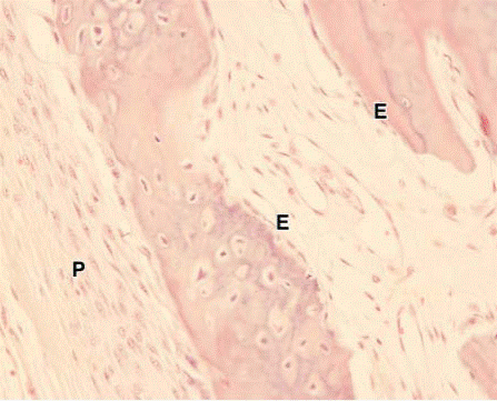
External and internal surfaces of bone are covered by layers of bone-forming cells and vascularized connective tissue called periosteum and endosteum.
The periosteum consists of a dense fibrous outer layer of collagen bundles and fibroblasts. Bundles of periosteal collagen fibers, called perforating(or Sharpey's) fibers, penetrate the bone matrix, binding the periosteum to bone. The innermost cellular layer of the periosteum contains mesenchymal stem cells called osteoprogenitor cells, with the potential to divide by mitosis and differentiate into osteoblasts. Osteoprogenitor cells play a prominent role in bone growth and repair.

Дата добавления: 2015-10-30; просмотров: 141 | Нарушение авторских прав
| <== предыдущая страница | | | следующая страница ==> |
| Fibrocartilage. | | | Periosteum and endosteum. |