|
Читайте также: |
CYTOLOGY AND EMBRYOLOGY
CELL
CELL STRUCTURE
| CELL | |
| CELL MEMBRANE (PLASMA MEMBRANE) | |
| - lipid - protein - carbohydrate | |
| PROTOPLASM | |
| - nucleus | Central more dense part of protoplasm. |
| 1. nucleoli | |
| 2. nucleoplasm | |
| 3. nuclear membrane | |
| 4. chromatin | |
| - cytoplasm | Outer less dense part of protoplasm. |
| 1. cytosol (hyaloplasm) | |
| 2. organelles | |
| - membranous organelles | Endoplasmic reticulum, Golgi complex, mitochondria, lysosomes. |
| - nonmembranous organelles | Cytoskeleton, centrioles, ribosomes. |


The fluid mosaic model of membrane structure.
(a): The fluid mosaic model emphasizes that a membrane consisting of a phospholipid bilayer also contains proteins inserted in it or bound to the cytoplasmic surface (peripheral proteins) and that many of these proteins move within the fluid lipid phase. Integral proteins are firmly embedded in the lipid layers. Other proteins completely span the bilayer and are called transmembrane proteins. Hydrophobic amino acids of the integral membrane protein interact with the hydrophobic fatty acid portions of the membrane. Both the proteins and lipids may have externally exposed oligosaccharide chains.
(b): Membrane splitting occurs along the line of weakness formed by the fatty acid tails of membrane phospholipids, since only weak hydrophobic interactions bind the halves of the membrane along this line. Electron microscopy of cryofracture preparation replicas is a useful method of studying membranous structures. Most of the protruding membrane particles seen (1) are proteins or aggregates of proteins that remain attached to the half of the membrane adjacent to the cytoplasm (the P or protoplasmic face). Fewer particles are found attached to the outer half of the membrane (E or extracellular face). For every protein particle that bulges on one surface, a corresponding depression (2) appears in the opposite surface.
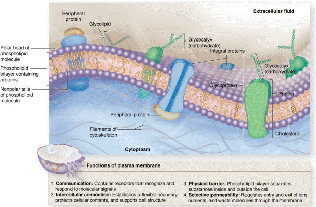
Membrane proteins.
Schematic drawing of plasma membrane structure shows a globular peripheral protein on the external face of the membrane and two integral transmembrane proteins.
One-pass transmembrane proteins have single hydrophobic regions along the length of amino acids and for maximal stability this becomes buried in the internal region of the lipid bilayer. Multipass transmembrane proteins have several hydrophobic amino acid sequences all buried in the bilayer, with terminal and intervening hydrophilic sequences exposed at either the external or cytoplasmic face of the membrane. Many physiologically important membrane proteins, including ion pumps and channels, are multipass proteins.
Proteins, which are a major molecular constituent of membranes can be divided into two groups. Integral proteins are directly incorporated within the lipid bilayer itself, whereas peripheral proteins exhibit a looser association with one of the two membrane surfaces. Some integral proteins span the membrane one or more times, from one side to the other. Accordingly, they are called one-pass or multipass transmembrane proteins.
Дата добавления: 2015-10-30; просмотров: 113 | Нарушение авторских прав
| <== предыдущая страница | | | следующая страница ==> |
| Two-cell stage | | | SIMPLE SQUAMOUS EPITHELIUM |