Though only snapshots of this highly dynamic organelle, electron micrographs of the Golgi apparatus provided early evidence about how this organelle functions, evidence that has now been strengthened by biochemical and other studies. To the right is a cisterna (arrow) of the rough ER containing granular material. Close to it are small vesicles containing apparently similar material. These are very close to the cis face of the Golgi apparatus. In the center are the characteristic flattened, curved, and stacked medial cisternae of the complex. Dilatations (upper left arrow) are seen extending from the ends of the cisternae. Similar dilatations gradually detach themselves from the cisternae and fuse at the trans face, forming the secretory granules (1, 2, and 3). Near the plasma membranes of two neighboring cells is more rough ER and smooth ER.
X30,000. Inset: a small region of a Golgi apparatus in a 1- m section impregnated with silver, which demonstrates the abundance of glycoproteins within some cisternae. X1200
Lysosomes

Lysosomes are large, generally spherical membrane-enclosed vesicles that function as sites of intracellular digestion and are particularly numerous in cells active in various
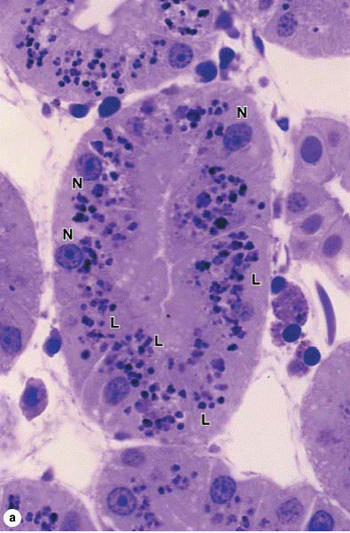
types of endocytosis. Lysosomes are not well-shown on H&E stained cells, but can be visualized by light microscopy after staining with toluidine blue. (a): Cells in a kidney
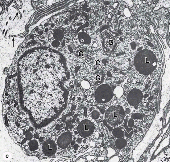
tubule show numerous purple lysosomes (L) in the cytoplasmic area between the basally located nuclei (N) and apical ends of the cells at the center of the tubule. Using endocytosis, these cells actively take up small proteins in the lumen of the tubule, degrade the proteins in lysosomes, and then release the resulting amino acids for reuse. X300 toluidine blue. (b): Lysosomes in cultured vascular endothelial cells can be specifically stained using fluorescent dyes sequestered into these organelles (green), which are abundant around the blue Hoechst-stained nucleus. Mitochondria (red) are scattered among the lysosomes. (c): In the TEM lysosomes (L) have a characteristic very electron-dense appearance and are shown here near groups of Golgi cisternae (G) and a centriole (C). Less electron-dense lysosomes represent heterolysosomes in which digestion of the contents is underway. The cell is a macrophage with numerous fine cytoplasmic extensions (arrows). X15,000.
Lysosomal hydrolases are synthesized and segregated in the RER and subsequently transferred to the Golgi apparatus, where the enzymes are further modified and ackaged in vacuoles that form lysosomes. The marker mannose-6-phosphate (M6P) is added by a phosphotransferase in the cis Golgi only to the N-linked ligosaccharides of the hydrolases destined for lysosomes. Membrane receptors for M6P-containing proteins in the trans Golgi network then bind these proteins and ivert them from the main secretory pathway for segregation into lysosomes.
Material taken from the cellular environment by endocytosis is digested when lysosomes fuse with the membrane of the phagosome or pinocytotic vesicle. The ndocytosed material mixes with the hydrolytic enzymes, a proton pump in the lysosomal membrane is activated to lower the internal pH, and digestion follows. The omposite structure is now termed a secondary or heterolysosome. Heterolysosomes are generally 0.2–2 m in diameter and present a heterogeneous appearance in he TEM because of the wide variety of materials they may be digesting.
During this digestion of macromolecules, released nutrients diffuse into the cytosol through the lysosomal membrane. Indigestible material is retained within the acuoles, which are now called residual bodies or telolysosomes. In some long-lived cells (eg, neurons, heart muscle), residual bodies can accumulate and are referred to as lipofuscin granules.
Дата добавления: 2015-10-30; просмотров: 116 | Нарушение авторских прав
| <== предыдущая страница | | | следующая страница ==> |
| Structural components of the nucleus. | | | Microtubules, cilia, and centrioles. |