|
Читайте также: |
CYTOLOGY AND EMBRYOLOGY
CELL
Cell is the fundamental unit of which all organisms are composed.
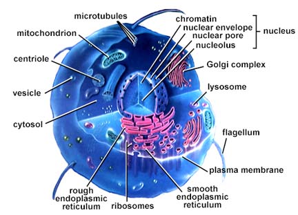
| CELL | |
| CELL MEMBRANE (PLASMA MEMBRANE) | The trilaminar membrane separating the cytoplasm of the cell from surrounding structutres. Plasmolemma is 8 to 11 nm thick. |
| - Lipids | |
| - Proteins | |
| - Carbohydrates | |
| PROTOPLASM | |
| - Nucleus | Central more dense part of protoplasm. |
| 1. nucleolus | A dense spherical accumulation of fibres and granules found in the nucleus. |
| 2. nucleoplasm | Nuclear sap which contains dispersed chromatin, some small granules, and protein. |
| 3. nuclear membrane | A boundary between the nucleus and the cytoplasm, composed of two unit membranes separated by the perinuclear space. At several points the inner and outer layers of the nuclear membrane fuse leaving gaps (nuclear pores). |
| 4. chromatin | Nuclear material that contains DNA and proteins. |
| - Cytoplasm | Outer less dense part of protoplasm. |
| 1. cytosol (hyaloplasm) | A fluid base or matrix of cytoplasm. |
| 2. organelles: | Intracellular structures having a specialized function. |
| - membranous organelles | Endoplasmic reticulum, Golgi complex, mitochondria, lysosomes. |
| - nonmembranous organelles | Cytoskeleton, centrioles, ribosomes. |

CELL MEMBRANE
LIPIDS. The trilaminar structure of cell membrane is produced by the arrangement of lipid molecules (predominantly phospholipids) that constitute the basic framework of the membrane.
Each phospholipid molecule consists of an enlarged head in which the phosphate portion is located; and two thin tails consisting of fatty acids.

PROTEINS. The proteins are present in the form of irregularly rounded masses.
Most of them are embedded within the thickness of the membrane and partly project on one of its surfaces: either outer (outer membrane proteins) or inner (inner membrane proteins).

However, some proteins occupy the entire thickness of the membrane and may project out of both its surfaces (these are called transmembrane proteins).

CARBOHYDRATES. Сarbohydrates are present at the surface of the membrane. They are attached either to the proteins (forming glycoproteins) or to the lipids (forming glycolipids). The carbohydrate layer is specially well developed on the external surface of the plasma membrane forming the cell boundary (glycocalyx).

The fluid mosaic model of membrane structure.
(a): The fluid mosaic model emphasizes that a membrane consisting of a phospholipid bilayer also contains proteins inserted in it or bound to the cytoplasmic surface (peripheral proteins) and that many of these proteins move within the fluid lipid phase. Integral proteins are firmly embedded in the lipid layers. Other proteins completely span the bilayer and are called transmembrane proteins. Hydrophobic amino acids of the integral membrane protein interact with the hydrophobic fatty acid portions of the membrane. Both the proteins and lipids may have externally exposed oligosaccharide chains.
(b): Membrane splitting occurs along the line of weakness formed by the fatty acid tails of membrane phospholipids, since only weak hydrophobic interactions bind the halves of the membrane along this line. Electron microscopy of cryofracture preparation replicas is a useful method of studying membranous structures. Most of the protruding membrane particles seen (1) are proteins or aggregates of proteins that remain attached to the half of the membrane adjacent to the cytoplasm (the P or protoplasmic face). Fewer particles are found attached to the outer half of the membrane (E or extracellular face). For every protein particle that bulges on one surface, a corresponding depression (2) appears in the opposite surface.

Дата добавления: 2015-10-30; просмотров: 132 | Нарушение авторских прав
| <== предыдущая страница | | | следующая страница ==> |
| Legal rights are, clearly, rights which exist under the rules of legal systems. | | | Structural components of the nucleus. |