|
Читайте также: |
The synapse (Gr. synapsis, union) is responsible for the transmission of nerve impulses from neuron to another cell and insures that transmission is unidirectional.
Synapses are sites of functional contact between neurons or between neurons and other effector cells. The function of the synapse is to convert an electrical signal (impulse) from the presynaptic cell into a chemical signal that acts on the postsynaptic cell. Most synapses transmit information by releasing neurotransmitters during this signaling process. Neurotransmitters are chemicals that bind specific receptor proteins to either open or closed ion channels or initiate second-messenger cascades. A synapse has the following structure:
- Presynaptic axon terminal (terminal bouton) from which neurotransmitter is released,
- Postsynaptic cell membrane with receptors for the transmitter and ion channels or other mechanisms to initiate a new impulse,
- 20–30 nm wide intercellular space called the synaptic cleft separating the presynaptic and postsynaptic membranes.
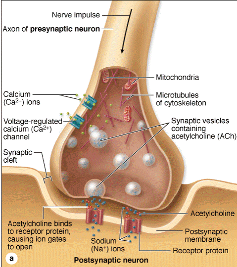
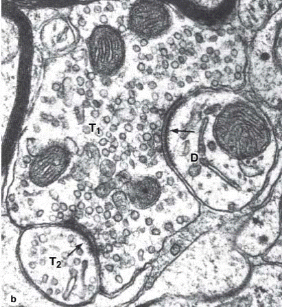
Synapse.
(a): Diagram showing how neurotransmitters are released from the terminal bouton in a chemical synapse. Presynaptic terminals always contain a large number of synaptic vesicles containing neurotransmitters, numerous mitochondria, and smooth ER as a source of new membrane. Some neurotransmitters are synthesized in the cell body and then transported in vesicles to the presynaptic terminal. Upon arrival of a nerve impulse, voltage-regulated Ca2+ channels permit Ca2+ entry, which triggers exocytosis releasing neurotransmitter into the synaptic cleft. Excess membrane accumulating at the presynaptic region as a result of exocytosis is recycled by clathrinmediated endocytosis, which is not depicted here. The retrieved membrane fuses with the SER in the presynaptic compartment for reuse in the formation of more synaptic vesicles. Some neurotransmitters are synthesized in the presynaptic compartment, using enzymes and precursors brought there by axonal transport. (b): TEM shows a large presynaptic terminal (T1) filled with synaptic vesicles and asymmetric electron-dense regions around 20–30 nm wide synaptic clefts (arrows). The postsynaptic membrane contains the neurotransmitter receptors and mechanisms to initiate an impulse at the postsynaptic neuron. The postsynaptic membrane on the right is part of a dendrite (D), associated with fewer vesicles of any kind, showing this to be an axodendritic synapse. On the left is another presynaptic terminal (T2), suggesting an axoaxonic synapse with a role in modulating activity of the other terminal. X35,000.
Nerve impulses sweep rapidly (in milliseconds) along the axolemma as an explosive wave of electrical activity (depolarization). At the presynaptic region the nerve impulse briefly opens calcium channels, promoting a calcium influx that triggers the exocytosis of synaptic vesicles. The released neurotransmitters diffuse across the synaptic cleft and bind receptors at the postsynaptic region, promoting a transient electrical activity (depolarization) at the postsynaptic membrane. These synapses are called excitatory, because their activity promotes impulses in the postsynaptic cell membrane. In some synapses the neurotransmitter-receptor interaction has
an opposite effect, promoting membrane hyperpolarization with no transmission of the nerve impulse. These are called inhibitory synapses. Thus, synapses can excite or inhibit impulse transmission and thereby regulate nerve activity.
Once used, neurotransmitters are removed quickly by enzymatic breakdown, diffusion, or endocytosis mediated by specific receptors on the presynaptic membrane. This removal of neurotransmitters is functionally important because it prevents an undesirable sustained stimulation of the postsynaptic neuron.
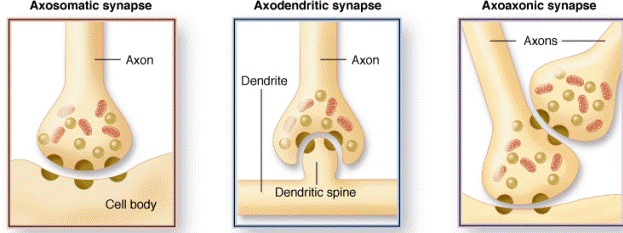
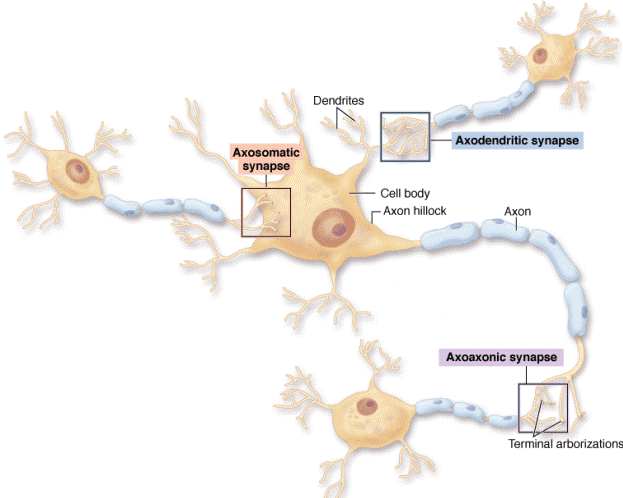
Morphologically, various types of synapses are seen between neurons. If an axon forms a synapse with a cell body, it is called an axosomatic synapse; with a dendrite, axodendritic; or with an axon, axoaxonic. The axoaxonic synapse is less common and is used to modulate synaptic activity.
Дата добавления: 2015-10-30; просмотров: 375 | Нарушение авторских прав
| <== предыдущая страница | | | следующая страница ==> |
| Dendrites and dendritic spines. | | | Oligodendrocytes |