|
Читайте также: |
The basic units of any device are the detector, the analyzer and the block of registration.
Clinical radionuclide evaluations are performed with the help of special devices. According to medico-functional purposes the radio diagnosis devices are divided into 3 groups.
1. Radiometers are used for measurement accumulation of γ - and β - radiating RPhP into the organs and tissues (DCU 2-1, GTRM-01, URZ-2, NP-354), body counter (body counting indicator) of a ionizing radiation in vivo and registration of the radionuclides contents into biological tests in vitro (URN-7, gamma-12, beta-2, Wallac).
2. Radiographs are applied for evaluation of timing characteristic of RPhP accumulation or distribution in an organism (URI-1, UR 1-3, RKA 3-01, KP-RDD-3 and others).
3. Gamma–topographers (gamma-ray irradiation device) are used for evaluation of spatial characteristics of RPhP distribution into the organism of the patient and obtaining of the two-dimensional image. Devices with the mobile detector (scintiscanner and gamma-ray chamber) allow to obtain of a gamma-topographical image of RPhP distribution into the body.
The single-photon emission computer tomography (SPECT) is a variant of gamma-ray chamber, which allows to receive the level-by-level scintigraphy images of RPhP distribution into the body.
The dates can be performed by different ways:
1) measurement of absolute or average quantity of impulses (with the radiometers);
2) making graphical model of time change of radioactivity (with the chronographs);
3) obtaining the picture of spatial distribution of RPhP into the body (with the scintiscanners, gamma-ray chambers, SPECT and PET).
As well all methods of nuclear imaging are divided into 2 big groups: evaluating the whole organism (in vivo) and evaluating the biological tests (in vitro).
Nuclear imaging in vivo:
Clinical radiometry - used to study relatively static, that is slow processes of accumulation and excretion of radioactive substances in the body and tissues when a one-shot measurement or re-measurement have taken a large interval of time– tens of minutes, hours, days. The sketch of radiometry see fig. 9.3.

Fig.9.3. The sketch of radiometric evaluation.
1 – Scintillation counter; 2 – the registering device.
Results of radiometry are expressed in percentage as relation the activity of radioactive substance which accepted by area of pathology by the activity of healthy tissues. Evaluation is fulfilled on the devices – radiometers ("Gamma", DCU-2-І, UR-3-2, etc.), and the results is received in the numerical value - Becquerel (Bk) – intensity of radiation from RPhP.
The scintillational detectors (so-called well counters) are used for determination of radio-activity of biological tests. Sketch of well counter see fig.9.4.
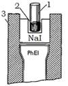
Fig.9.4. Sketch of well counter. 1 – test tube; 2 – radioactive sample; 3 – leaden collimator; NaI – scintillator; PhEI – photoelectronic increaser.
Determination of radioactive sample activity: the test tube with sample put into a well counter. The radiation of radioactive test works up scintillator and is cause the flashes of light in it. The flashes of light with the help of photoelectronic increaser transform in the electrical impulses. Results are obtained as a numerical value of activity in Bk.
Radiography is used to study rapid physiological processes - circulation time test, ventilatory capacity (ventilating function) of lungs, a functional state (functional condition) of heart, liver, kidneys, etc. Results are obtained as graphical model (curves of radio-activity), which show of time change of radioactivity in the selected area. The basic sketch the radiographs see fig.9.5.

Fig.9.5. Sketch of radiographic evaluation.
1 – Scintillation counter; 2 – a control panel; 3 – a recorder.
Scanners are used for Scanning. The scanner is the device with movable detector (a crystal potassium iodide in diameter 50 mm) and the device for creation of the image that is for visualization of distribution RPhP in organism. The registration (in the form of line mark) is performed gradually – field to field. Distribution of RPhP is estimated according to opposition and density of line mark: in those places where the density of line marks is greater, the quantity of RPhP is more. The image is named scanning. The basic sketch of scanning see fig.9.6.
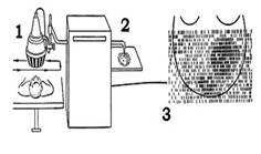
Fig.9.6. the basic sketch of the scanner.
1 – Scintillation counter; 2 – a recorder; 3 – scanogram of abdominal cavity.
Scintigraphy it is carried out by means of gamma-ray chamber. The gamma-ray chamber is the radio-diagnosis device; the main part being the big motionless detector – a monocrystal potassium iodide in diameter 40-60 cm with plenty of photo-electronic multipliers (PEM) located on it. They transform flashes of light on the whole surface of a monocrystal into electric impulses and after their processing by a computer; the image arises on the screen of the monitor. Thus the distribution of flashes to the screen beats off distribution of scintillation in a crystal, and they in turn reflect a picture of distribution and speed of moving of the gamma-radiating RPhP in the body. Advantage of the gamma-ray chamber as compared to the scanner is that gamma-ray chambers allow us to receive simultaneously information about distribution of RPhP in the entire body and to investigate rapid physiological processes (a blood-stream in the organ, distribution of radioactive gas 133Хе in alveoli of lungs at breath) by viewing the screen, video magnetic record. The basic sketch of work of gamma -chamber see fig. 9.7., 9.8.

Fig.9.7.The basic sketch gamma–ray chamber: 1- detector; 2-control panel; 3-monitor; 4-scintigramm of lungs

Fig.9.8 The sketch of the detector gamma-ray chambers: 1-collimator; 2-scintillation detector; 3- optical waveguide; 4-the electronic scheme; 5-lead shielding; Ph - photomultiplier tube.
Дата добавления: 2015-10-23; просмотров: 182 | Нарушение авторских прав
| <== предыдущая страница | | | следующая страница ==> |
| Pharmaceutical requirements for RPhP | | | Radionuclide computer tomography includes a single-photon emission computer tomography(SPECT) and two-photon positron emission tomography(PET). |