Читайте также:
|
Cyanide poisoning caused by nitroprusside n house fires,
Rx. Amil nitrate which forms METHb that combines to CN followed by thiosulphate which then forms thiocyanate!!!
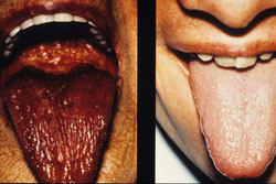
| 1. Patient: central cyanosis The patient on the left has a markedly cyanotic (blue) tongue compared to the normal patient�s tongue on the right. Central cyanosis is due to an increased amount of unoxygenated hemoglobin (Hgb) in the blood (called deoxyhemoglobin). This translates into a decrease in the oxygen saturation of blood (percentage of the heme groups on Hgb occupied by oxygen). Central cyanosis is due to any cause of hypoxemia (e.g., respiratory acidosis, ventilation/perfusion/diffusion problems in the lungs, right to left shunting of blood in cyanotic congenital heart disease), since a low arterial PO2 automatically lowers the amount of O2 that diffuses into the RBC to bind to heme. Peripheral cyanosis refers to vasoconstriction of the blood vessels in the skin with central shunting of the blood (e.g., hypothermia). The skin is cyanotic, however the mucous membranes are pink. When Sao2 < 80, cyanosis becomes visible!! |
O2 Hb Dissociation Curve: “S” shaped. At PaO2 of 25-27 mmhg (P50), saturation is 50% ---- at 40mmhg it goes upto 75% ---- and at 100 mm-hg it goes to 95% which is the o2 carrying capacity of Hb!!
Shifts: key “LA,RD” left à Hb association with o2
Right à Hb dissociates with o2.
Right shift O2 Curve: decreased affinity to O2
Increase PaCO2, decrease PaO2,
BPG Increase,
High altitude,
4. Acidosis(increased H+ concentration),
5. Fever.
Left Shift O2 Curve: increased affinity to O2
CO,
MetHb,
HbF;
BBG dec,
5. Alkalosis
Cytochrome oxidase delivers electron to O2 --- Inhibited by CO n Cyanide (3 “C”)
Uncoupling Agents (inner Mitochondrial Membrane permeable to protons): Alcohol, salicylates,dinitrophenol(chemical used to preserve wood).
Failing of NADH, FAD producing systems (which produce protons which leak through?? Actually leaking of prons causes failure of those systems..) ---> so where would the energy go if not into ATP?? --->heat causes hyperthermia.
Alcoholics ---> Uncoupling Agent ---> Heatstrokes risk increase
Arterio-venous shunting: may also cause hypoxia
1. AV fistula from trauma: bypass from microcirculation
2. Spider angiomas: due to hyperestrinism??
3. Mosaic bone in paget’s disease of bone!!
Resp Acidosis --- Hb Normal, O2 Sat dec, Po2 dec
Anemia --- Hb decrease, Saturation Normal, Po2 Normal
Co+ MetHb --- Hb Normal, Saturation dec, Po2 normal
hypoxia à ATP depletion
--> Anaerobic Glycolysis is activated by Phosphofructokinase due to low levels of citrate n AMP--- all cells can do including. RBC
---> NADH is converted into NAD+ and pyruvate is converted into lactate!! NAD+ is used to produce 2 ATPs per glucose
---> Increase of lactic acidosis (anion gap acidosis???)
---> acid raised
---> protien denaturation ---> enzymes denatured as well ---> called coagulation necrosis.
Therefore Tissue hypoxia ---> Acidosis ---> coagulation necrosis ultimately
Anaerobic Glycolysis ---> screws all the ATPase pumps ---> esp. Na + Water enter cells
---> cell swelling first histologic sign of reversible cell injury
Ribosomes fall off from endoplasmic reticulum!! Decrease in protein synthesis!!
Digitalis blocks na/k ATPase pump à influx of Na+ à release of Ca+2 à contractility increased!!
No ATP ---> irreversible cell damage ---> Ca-Mg ATPase pump dec ---> Intracellular Ca increase ---> Mitochondrial enzymes activated like phospholipase lead to Membrane Damage ---> nuclear enzymes, endonuclease leads to pyknosis ---> Phospholipase damage to cell membrane ---> Pancreatitis.
Cell death: CK-MB, troponins I in heart, SGOT, ALT in hepatitis, Amylase in acute pancreatitis increase when cell dies..
Lipofusin is end product of lipid peroxidation---> when free radical damage ---> wear n tear pigment ---> brown pigment ---> atrophic organs in elderly n cancer patients brown atrophy---> pigment cant be digested.

| 2. Liver biopsy: lipofuscin The brown pigment noted in the hepatocytes (arrow) represents lipofuscin (wear and tear pigment). Lipofuscin is the end product of free radical injury of cells and autophagy (destruction of cell organelles and structural proteins in cells undergoing atrophy). Grossly, an excess of lipofuscin produces a brown discoloration, hence the term brown atrophy. Lipofuscin is commonly found at autopsy in elderly patients. |
O2 dependant Myeloperoxidase system (MPO):
Most lethal bactericidal system present in neutrophils n monocytes!! (not in macrophages)
NADPH oxidase forms molecular O2 in Superoxide free radical which is very toxic!!
Reperfusion injury is due to O2 Radicals e.g. TPA in thrombotic artery cause lysis n blood supply with o2 which may cause injury due to formation of free radicals..
O2 Radical ---> destroys retina ---> blindness. Retrolental dysplasia!!
O2 Radicals ---> Bronchopulmonary dysplasia (fibrosis)
Ionizing radiation to treat cancerà formation of hydroxyl radicals from water à causes gene mutation à may cause cancer (MC= Leukemia)
Iron loves making free radicals….
Fenton reaction: H2O2 + fe2 à fe3 +OH- + OH ----> cirrhosis, restrictive cardiomyopathy, pancreastic failure ---> Malabsorption n diabetes.
------------------------x--------------------x------------------------
peroxide is neutralized by catalase n glutathione!!
Tylenol (Acetaminophen) ---> produce free radicals ---> #1 cause of drug induced Fulminant hepatitis (around central vein) ---> kidney damage ---->
Rx: N-Acetyle Cystein (Increases Glutathione which neutralizes free radicals)
Superoxide dismutase neutralize superoxide ---> producing peroxide
methotrexate causes BM suppression so we give leucovarine to prevent the side effect!!
Pentose/hexose phosphate shunt makes Glutathione and NADPH
CCl4(dry cleaning) converted in to CCL3 ---> fulminant hepatitis
Acetaminophin (Tylenol) + Aspirin ---> damages kidney big time (renal medulla) (pappilary necrosis??)----> PG e2 dec
Apoptosis ---> genes involved in prog cell death ---> (eg. embryo)
Very important in embryology!!
how does the solid bowel becomes tubular?? Due to apoptosis
King of our body ---> Y chromosome ---> Mullerian inhibitory factors(signal) ---> erase mullerian str(uterus, cervix, upper third of vagina) ---> caspases wipe them ---> Apoptosis ---> producing lipofuscin and apoptotic bodies!!
Cell Injury
Post 3: (Dated: Jan 13 - 2009)
In women ---> X chromosome ---> wolfian duct structures(epidydimus, seminal vesical, vas differense) are removed by apoptosis
Anterior mediastenum is smaller in adults as compare to children (Due to thymus) ---> apoptosis in adults
Digeorge syndrome ---> Thymus absent ---> classic t cell def -->cause hypocalcemic tetany due failure of parathyroid.
Apoptosis occurs in cancer cells
Apoptotis occurs in neuron cells ---> dec in brain mass(atrophy) ---> pyknotic bodies.
Caspases are involved in Apoptosis
Apoptosis in viral hepatitis induced by CD8 T cells leaving councilman bodies!!

| 3. Liver: viral hepatitis with apoptosis The arrow points to an anucleate, deeply eosinophilic staining cell that is retracted away from the other cells. The last phase of destruction of this cell will be fragmentation into apoptotic bodies containing bound packets of organelles which will be phagocytosed and destroyed in the lysosomes of macrophages and neighboring cells. Note that there is no inflammatory infiltrate around the cell. (Ci202) |
Necrosis

| 5. Heart: coagulation necrosis Recall that in coagulation necrosis, the accumulation of lactic acid in the hypoxic tissue causes denaturation of the proteins and enzymes in the cell. Hence, the cells retain their basic outline but cannot be autodigested owing to the denaturation of their enzymes. Neutrophils from outside the area of infarction will have to destroy the tissue, which will eventually be replaced by collagen in a scar. Note that the cells are deeply eosinophilic (see arrow). Nuclei and cross striations are not evident. |
ischemia--> inc lactic acid --> denatures proteins n enzymes -->Coagulation necrosis (micro)---> (grossly)Pale infarct in tissues with good consistency so that RBCs don’t get out of vessels ---> looks pale - Heart, kidney, spleen(usually) n liver(rare coz of the double blood supply)
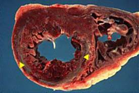
| 4. Heart: pale infarction with coagulation necrosis The anterior part of the heart is at the bottom of the slide. The left ventricle is on your left and the right ventricle on your right. The arrows point to a pale infarct in the entire anterior part of the heart and anterior two-thirds of the interventricular septum. The patient had a platelet thrombus in the left anterior descending coronary artery, which supplies blood in the same distribution outlined by the infarction. Coagulation necrosis is the underlying pathologic process. The infarct is pale owing to the dense consistency of muscle so that extravasated RBCs from necrotic vessels remain localized and are unable to percolate through the tissue. | |
Hemorrhagic infart in tissues with loose consistency so that RBCs get out of vessels n looks hemorrhagic: Bowel, testicle (torsion), lungs
coagulation necrosis depends on consistency of tissue. Either it becomes pale infarct or hemorrhagic infarct.
Left side of heart ---> most of the emboli arises
vegetation of acute rheumatic fever rarely embolize ---> Infective endocarditis does produces them mostly
Clots of emboli or vegetation goes to spleen causing pale infarction
The arrhythmia mostly associated with systemic emboli is atrial fibrillation ---> causes stasis, clot formation n vibration so that clots can detach n become emboli!!
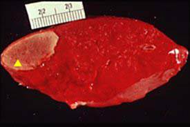
| 7. Spleen: pale infarction Note the roughly wedge shaped pale area of infarction extending to the surface of the splenic capsule (see arrow). The infarct is pale since the tissue texture of the spleen does not allow RBCs to diffuse easily through the tissue.Most causes of splenic infarctions originate from embolization of clot material or bacterial vegetations from the left heart into the aorta with eventual deposition in distal organs. If the splenic vein was thrombosed, the infarct would have been hemorrhagic. |
Dry gangrene: no pus ---> Diabetics’ foot
Most common cause of non traumatic amputation is diabetes ("ATHerosclerosis of popliteal artery)
Popliteal artery a dangerous artery just after coronary.

| 6. Toe: dry gangrene in a patient with diabetes mellitus (coagulation necrosis)The big and little toe are blackened. A pressure ulcer is noted overlying the metatarsal head of the big toe. The cause of the gangrene is artherosclerotic occlusion of the popliteal artery. This is a dry gangrene with predominantly coagulation necrosis since there is no evidence of exudate indicating an anaerobic infection. In the latter case, the predominant type of necrosis is liquefactive necrosis, since infection with neutrophils is present. This is called wet gangrene. (Ci206) |
Infection superimposed on dry gangrene = wet gangrene!
Most common cause of bowel infarction(hemorrhagic): #1: adhesions of previous surgery, #2: piece of small bowel trapped in an Indirect inguinal hernia in a sac.
Lung: Hemorrhagic infarction(due to pulmonary emboli) ---> wedge shape ---> effusion present --> neutrophils present ---> pluritic chest pain
| 9. Lung: gross of a pulmonary infarction (hemorrhagic infarct) Note the wedge-shaped area of hemorrhage extending to the pleural surface (see arrow). This is an example of a hemorrhagic infarction and is most often secondary to emboli originating from clots that developed in the femoral veins in the legs. Most venous clots initially develop in the deep veins of the calf, however, as they propagate proximally into the femoral venous system, they break off and embolize to the lungs. The embolized vessel is visible at the apex of the infarct (see arrow). Recall that hemorrhagic infarcts usually develop in tissues that have a loose texture, so that blood extravasating from necrotic vessels can percolate through the tissue. The wedge-shaped infarct is due to the parallel branching of the pulmonary vessels. Hence, a proximally located obstruction in the lumen of a pulmonary vessel affects the blood flow to a large cross-sectional area of branching vessels that are distal to the obstruction (perfusion defect). The same concept applies to a mucous plug in a terminal bronchiole. Recall that the small airways also branch in parallel, hence a proximally located plug affects a large cross-sectional area of distally branching airways. |
Liquefactive necrosis (in brain due absence of fibroblasts and presense of astrocytes which provides the holding working like fibroblasts,but they cant produce collagen so here it would be liquefactive necrosis with cyst formation rather than coagulative even though the cause is ischemia)

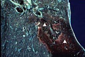
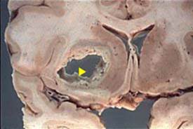
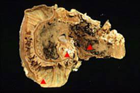
| 10. Brain: old artherosclerotic stroke with cystic cavity (liquefactive necrosis)Note the cystic cavities in the brain. Recall that infarction in the brain is not coagulative but liquefactive necrosis owing to the lack of good structure in the brain and the abundance of lysosomes in neuroglial cells. |
Liquefactive necrosis is related to an infection - neutrophils present - cyctic necrosis - acute inflamation related to neutrophils - damaging the tissues --> may produce abscess.
| 11. Brain: cerebral abscess Note the cavitary lesion in the brain, which is lined by a shaggy, necrotic material (see arrow). Cerebral abscesses, like cerebral infarctions, are examples of liquefactive necrosis. Cerebral abscesses may be due to local extension of an infection (e.g., mastoiditis, sinusitis) or hematogenous spread of a bacteria. |
Gram + cocci in clusters = staph aureas ---> has coagulase --> converts fibrinogen into fibrin --> localize inf by restricting wbc --> abcess
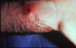
| 12. Wrist: abscess due to Staphylococcus aureus (liquefactive necrosis)Note the raised, erythematous area on the wrist with a central area of draining from a sinus tract. Recall that S. aureus is a gram positive coccus that has coagulase. Coagulase converts fibrinogen into fibrin, which keeps the infection localized. This is an example of liquefactive necrosis. |
strept realeases hyaluronidase--->breakdowns glycosaminoglycans (conn tissue) ---> cellulitis ---> infetion spreads thru tissues
which type of necrosis is asso with inf n neutrophils?--> liquefactive necrosis
yellow areas in lung in patient with high fever n productive cough --> gram stain?? --> gram + diplococci in chains-->strep pneumonia
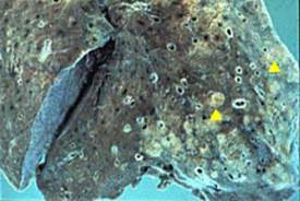
| 13. Lung: gross of lung exhibiting bronchopneumonia Note the patchy yellow, raised areas throughout the lung parenchyma (see arrow). These represent collections of neutrophils in the pulmonary parenchyma that first caused acute inflammation involving the small airways (acute bronchitis) and spilled over into the surrounding lung parenchyma causing bronchopneumonia. The progression of a bronchitis into the lung parenchyma is called bronchopneumonia. Streptococcus pneumoniae, a gram positive diplococcus, is the most common pathogen responsible for community acquired bronchopneumonia. Bronchopneumonia may cause liquefactive necrosis, since neutrophils are present in the tissue and producing enzymes which may destroy the tissue. |
Granulomatos necrosis ---> “caseous” ---> mycobacteria or systemic fungal infection ---> lipid in cell wall giving cheasy appearance
Non caseous granulomatous ---> Sarcoidosis n crohn’s disease
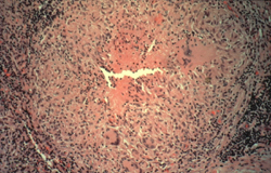
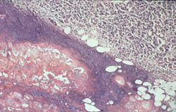
| 14. Lymph node: localized granuloma (caseous necrosis) in a patient with tuberculosis Note the very well circumscribed eosinophilic staining granuloma. In the middle of the granuloma, the pink staining material is acellular. This represents caseous necrosis. Note the multinucleated giant cells at 2 and 7 o'clock. The following slide is a high power view of the granuloma. | |

| 15. Lymph node: localized granuloma (caseous necrosis)in a patient with tuberculosis Note the eosinophilic appearing necrotic center of the granuloma (see arrow) and the wall of the granuloma containing a multinucleated giant cell (see arrow). The pink staining cells in the wall represent activated macrophages (called epithelioid cells). The interaction of macrophages containing processed TB organisms causes the helper T cells to release gamma interferon, which activates the macrophage and allows it to kill the TB. The cell walls of TB are rich in lipid, which causes the necrotic debris to have a cheezy appearance, hence the term caseous necrosis. Multinucleated giant cells represent macrophages fused together. Granulomas are an example of type IV cellular immunity. |
Epigastic distress with pain radiating to back ---> pancreatitis (retroperitoneal) ---> Enzymatic fat necrosis --> chawky white areas (saponification)
Pendulous breast ---> Traumatic Fat Necrosis ---> Calcification ---> painful (not in cancer) ---> Xray looks like cancer ---> Calcium bound to fatty acid = saponification
Bluish discoloration ---> due to calcium ---> dystrophic calcification
Blue discloration with athromatous plaque = calcium
Lipases most specific for pancreatitis, amylase can also increase in parotid, small bowel or fallopian tubes!!
| 16. Pancreas: acute pancreatitis with enzymatic fat necrosis Note the C shaped pancreas on the left of the slide. The duodenum encompasses the head of the pancreas and the body and tail extend to the right. Note the chalky white areas (see arrows) representing calcium interacting with fatty acids derived from breakdown of fat by pancreatic lipases. This is called saponification. The calcification is also an example of dystrophic calcification, which is calcification of damaged tissue. Pancreatic calcifications are visible on x-ray, ultrasound, or CT scans. The red areas in the pancreas (see arrows) represent hemorrhage due to rupture of vessels destroyed by elastases in the pancreas. Acute pancreatitis is most often due to alcohol and biliary tract disease. Serum lipase and amylase are both elevated. Pain radiates into the back since the organ is retroperitoneal. (Ci010) | |
| 17. Pancreas: histologic section of enzymatic fat necrosis in an alcoholic with acute pancreatitis The blue staining area represents enzymatic fat necrosis where calcium has bound with fatty acids to form a calcium salt. These can be visualized on x-ray, particularly after repeated attacks of acute pancreatitis. Calcium deposits in necrotic tissue are called dystrophic calcification. |
Alcoholics --> pancrease big time damage
alcohol is the most common cause of fatty change and cirrhosis of the liver
Gummatous necrosis ---> unique for tertiary syphilis!!
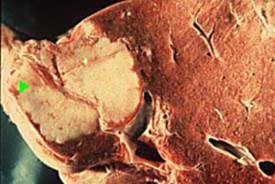
| 19. Liver: gumma in tertiary syphilis Note the white nodular lesions in the liver parenchyma (see arrow). Gummas have a rubbery consistency. They are examples of gummatous necrosis, which is unique to tertiary syphilis. There is nothing on gross examination of these masses that would distinguish them from primary or metastatic cancer, hence a microscopic section and history would be necessary to make the correct distinction. |
Fibrinoid necrosis ---> necrosis of immunologic disease (Rheumatic fever, RA(joint morning stiffness), glomerulonephritis, SLE, Vasculitis)
Palpable purpura ---> small vessel vasculitis ---> type 3 immune complex)hypersensitivity
Immune complex ---> activates alternative complement system ---> C5a ---> chemotaxis of neutrophils ---> cause actual damage
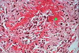
| 18. Arteriole: fibrinoid necrosis Note the pink staining material in the wall of this arteriole (see arrow) that has been totally destroyed. This is an example of small vessel vasculitis, the majority of which are due to type III immunocomplex disease.Immunocomplexes deposit in the vessel and activate the alternative complement system, which releases C5a, a chemotactic agent that attracts neutrophils. The neutrophils destroy the vessel. The pink staining material represents protein from the plasma in the vessel lumen. Fibrinoid necrosis is the necrosis of immune disease and is seen in the vegetations associated with acute rheumatic fever and inflammation associated with many collagen vascular diseases. |
portal triad = portal vein, hepatic artery n bile ducts
Sinusoidal organs ---> liver, spleen n bone marrow!!
Sinusoids = charaterstic gaps btw endothelial cells ---> large cells can pass through them v/s GBM where the cells have pores in them!!
Rt heart failure ---> backup of blood --->liver congestion “nutmeg” liver
Blockage of hepatic vein---> budd chiary syndrome --> liver congestion!!
Area around central vein of liver most susecptible to injury normaly ---> Zone 3 --> as being more hypoxic!!
Yellow fever (Arbovirus) damage the liver zone 2 (Mid-zone necrosis)
Zone 1 --> near the portal triad!!
Дата добавления: 2015-10-24; просмотров: 189 | Нарушение авторских прав
| <== предыдущая страница | | | следующая страница ==> |
| Companies Analysis | | | Restricting the carbohydrates will reduce the synthesis of VLDL |