|
Читайте также: |
MTX, used in rheumatoid arthritis, inhibits DHF reductase S phase---> can Cause macrocytic anemia(folate def).
Atrophy = decrease in tissue mass, decrease cell size, less mitochondria --- just have enuff organeles to survive.
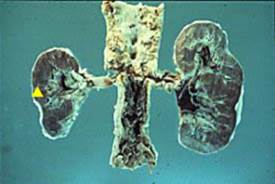
Hydronephrosis is mostly caused by renal stones ---> increase pressure in cortex n medulla(compression atrophy)---> ischemia ---> atrophy of renal tubules.
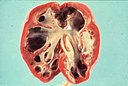
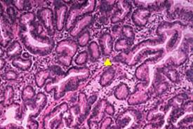
41. Kidney: hydronephrosis with compression atrophy
Note the dilated renal pelvis and calyceal system and the thinning of the renal cortex related to the increased pressure. The increased pressure imposed on the renal cortex causes it to undergo atrophy (compression atrophy). Hydronephrosis always implies obstruction to urine flow, which may be secondary to prostate hyperplasia, a renal stone (most common), pregnancy, or cancer at the ureterovesical junction.
ATH of cerebral artery, Alzheimer (β amyloid protein, in layers 3,5,6 destroyed) ---> brain atrophy, neuron degeneration ---> reduction in mass of brain
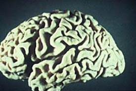
| 39. Brain: atrophy Note the loss of brain mass, which causes separation of the gyri and sulci. This is most commonly caused by atherosclerosis of the internal carotid artery. Chronic tissue hypoxia causes the neurons in layers 3, 5, and 6 of the cerebral cortex to undergoapoptosis. Drop out of these neurons causes a loss in brain mass, which is evident in this slide. Other causes include degeneration of the neurons in Alzheimer's disease or destruction of neurons by infection. |
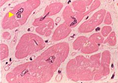
Lower motor neuron loss like ALS, lue garret disease??--> muscle not innervated -->undergoes atrophy!!
immobilization Casts --> decrease in muscle mass.
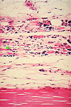
43. Muscle: atrophy
Note the markedly atrophic muscle fibers with eosinophilic cytoplasm and pyknotic nuclei (see arrow). Skeletal muscle atrophy may occur with disuse (e.g., cast on the leg), primary muscle disease (e.g., Duchenne's muscular dystrophy), or loss of nerve stimulation (e.g., amyotrophic lateral sclerosis, compression of a nerve by a ruptured intervertebral disc).
Hypopituitarism ---> adrenal gland atrophy (only zona reticularis-->sex hormones and fasiculata-->steroids)
not zona glomerulosa --> ACTH has nothing to do with aldosterone release!
ACTH responsible for glucocorticoid (Z. Fasciculata), and sex hormones (Z. Reticularis); Not aldosterone (zona glumerulosa)
Oral thyroid ---> thyroid stimulating hormone goes down ---> thyroid atrophy.
Cystic Fibrosis (defective cystic fibrosis transmembrane regulator at Ch.7) ---> problems with thick secretions of exocrine part ---> block lumen --->back pressure --> pancreas atrophy ---> malabsorption
| 44. Pancreas: cystic fibrosis (atrophy)Note the eosinophilic material blocking the ducts and the increase in connective tissue surrounding the ducts. Thick secretions in CF block the lumens and the pressure is transmitted back into the exocrine glands leading to atrophy via individual cell necrosis (apoptosis). Loss of exocrine secretions leads to malabsorption of fats, carbohydrates, and protein. |
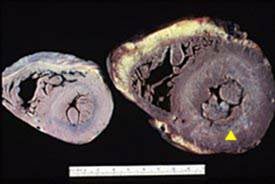
Atheromatous plaque in renal vessel --->Renal vascular hypertension ---> Renin high ---> diseased kidney get atrophied, other kidney hypertrophy.
Atrophy of one kidney will cause compensatory hypertrophy of the other!!
| 42. Kidneys, renal arteries, aorta: renal atrophy owing to atherosclerosis of the proximal portion of the renal artery The small kidney on your left (see arrow) has undergone atrophy owing to an atherosclerotic plaque partially blocking the opening of the renal artery. Note that the renal artery in the normal kidney is patent. Recall that reducing blood flow to the kidney leads to stimulation of the renin-angiotensin-aldosterone system. The juxtaglomerular apparatus located on the afferent arteriole is stimulated to release the enzyme (not hormone) renin. Renin cleaves angiotensinogen into AT-I. AT-I is converted into AT-II in the lungs by the enzyme, angiotensin converting enzyme (ACE). ATII is a potent vasoconstrictor and also stimulates the 18-hydroxylase enzyme in the zona glomerulosa to convert corticosterone into aldosterone. Aldosterone reabsorbs sodium in the distal and collecting tubules, hence increasing plasma volume. Hypertension, is the end-result of peripheral vasoconstriction of the arterioles by ATII and increased plasma volume by aldosterone. Hypertension secondary to renal artery atherosclerosis is called renovascular hypertension. As expected, the renal vein renin levels in the small kidney are increased. However, renal vein renin levels in the normal kidney should be suppressed, since the increased levels of ATII and aldosterone inhibit renin release from the normal kidney. |
If you block G2 phase, you have 4n chromosomes (no mitosis) ---> increase in size of cell (hypertrophy of cardiomyosite)---> copies of structures inside cells
1n - sperm
2n - Diploid cell
3n - Cancer cell or some trisomy disease
| 45. Hearts: normal on the left and concentrically hypertrophied on the right The heart on the left is normal. The heart on your right has left ventricular hypertrophy (LVH) with concentric hypertrophy of the muscle (see arrow) leading to a diminished left ventricular chamber with very little space for blood. Concentric hypertrophy of cardiac muscle is due to an increased resistance against which the ventricles must contract to expel blood out of the ventricle (increased afterload=pressure overload hypertrophy). Examples include the increased peripheral resistance in the arterioles associated with essential hypertension (most common) and aortic valve stenosis (narrow opening to the valve). | |
| 46. Muscle: hypertrophy Note the increased size of the individual muscle fibers and increased size of the nuclei representing hypertrophy (see arrow). Hypertrophy of a muscle increases the force of contraction of the muscle, since there are twice as many contractile fibers in the cytosol than a normal muscle. Recall the hypertrophied cells pass through the S phase but cannot undergo mitosis, hence they have twice the DNA (4N). |
if left unchecked, Hyperplasia is predisposing for cancer (EXCEPT prostate)
Unopposed estrogen (no progesterone) cause endometrial hyperplasia ---> atypical hyperplasia ---> endometrial cancer.
“progesterone undo estrogen”
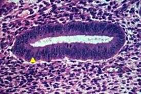
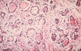
| 47. Endometrial mucosa: normal endometrial gland in the proliferative phase of the menstrual cycle Recall that the proliferative phase of the cycle is an estrogen primed event. Estrogen is a growth promoter and causes the endometrial glandular cells to mitose and undergo mitotic divisions (see arrow), which cause the glands to increase in number. This is an example of physiologic hyperplasia. | |
| 48. Endometrial mucosa:pathologic endometrial hyperplasia due to unopposed estrogen stimulation When comparing this slide with 047, it is apparent that far more glands are present and they are crowded together. This occurs when a woman has too much estrogen and not enough progesterone, the latter a hormone that causes endometrial glands to undergo atrophy. Examples of unopposed estrogen include a postmenopausal woman who is taking estrogen without progesterone to prevent osteoporosis or an obese woman, who has increased aromatization of androgens into estrogens in the adipose. If endometrial gland hyperplasia persists, endometrial dysplasia, and eventually endometrial carcinoma may occur. |
Дата добавления: 2015-10-24; просмотров: 99 | Нарушение авторских прав
| <== предыдущая страница | | | следующая страница ==> |
| Restricting the carbohydrates will reduce the synthesis of VLDL | | | Prostate hyperplasia does not predispose to cancer. |