Читайте также:
|
The developing embryoblasts induce changes in the endometrium in their vicinity, altering it to begin the formation of the material portion of the placenta. This altered maternal tissue, called the decidua, is subdivided into three regions:
1. The decidua capsularis.
2. The decidua basalis.
3. The decidua parietalis.
Figure 9. Schematic diagram of mature human placenta
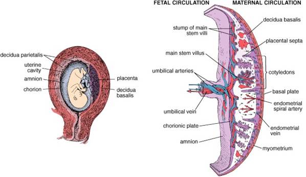
Figure10. Schematic diagram of sections through a developing human embryo at 16 days (a) and at 21 days (b) gestation

The region of the chorion in contact with the decidua capsularis forms short villi and known as the chorion laeve. The region of the chorionic plate in contact with the decidua basalis forms extensive chorionic villi, primary villi; this region is known as the chorion frondosum or villous chorion.
With further development, extraembryonic mesenchymal cells enter the core of the primary villi, converting them into secondary villi. The connective tissue of the secondary villi becomes vascularized by extensive capillary beds, which are linked to the developing vascular supply of the embryo. So appear tertiary villi. The decidua basalis forms large vascular spaces, lacunae that are compartmentalized into small regions by placental septa, extensions of the decidua. The fetal part of the placenta is divided by placental septa into 15 areas called cotyledons. Wedge-like placental septa form the boundaries of the cotyledons, and because they do not fuse with the chorionic plate, maternal blood can circulate easily between them.
Cotyledons are visible as the bulging areas on the maternal side of the basal plate. Fetal blood enters the placenta through a pair of umbilical arteries. Fetal blood returns through a system of veins that parallel the arteries except that they con-verge on a single umbilical vein.
Secondary villi project into these vascular spaces and are surrounded by maternal blood that is delivered to and drained from the lacunae by maternal blood vessels of the decidua basalis. Most of the villi are not anchored to the decidua basalis but are suspended in maternal blood of the lacunae; these are known as free villi. The villi anchored to the decidua basalis are called anchoring villi. Capillaries of freeand anchoring villiare near the surface of the villi and are separated from the maternal blood by a slight amount of connective tissue and the syncytiotrophoblasts covering the secondary villus.Thus, maternal blood and fetal blood do not intermix; instead, nutrients and oxygen from the maternal blood diffuse through the syncytiotrophoblasts, connective tissue, and endothelial cells of the capillaries of the villito reach the fetal blood.
Дата добавления: 2015-07-25; просмотров: 244 | Нарушение авторских прав
| <== предыдущая страница | | | следующая страница ==> |
| Implantation | | | UMBILICAL CORD |