Читайте также:
|
The device forSPECT represents a gamma-chamber in which during evaluation of the patient the detector moves around the investigated part of the body. To carry out the tomography gamma-radiating RPhP, one photon is emitted at single disintegration.
The level-by-level images are reconstructed by means of computer. SPECT has high resolution in comparison with plane scintigraphy. Appearance of a single-photon emission computer tomography see fig. 9.9.
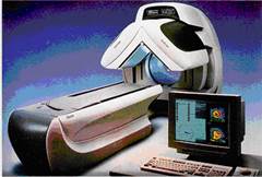
Fig.9.9. Appearance of SPECT.
Positron emission tomography (PET) - method of evaluating the functional condition of the tissues and organs with positrons. Taking off from an atom, each positron cooperates with an electron, therefore annihilation occurs - both particles disappear, and instead of them, two quanta arise and then scatter in epifocal directions in a positron tomography at the level of an investigated part of a body of the patient. 2 detectors are placed which move in a circle.Simultaneous registration of two - quanta which have arisen during annihilation, testifies to destruction of a positron on a line which connects 2 points of detection. Basic sketch of PET see fig. 9.10. Appearance of PET see fig. 9.11.

Fig.9.10 A principle of a positron emission tomography
 The annulations of positron with electron with two photons formation, moving in oppose-focal directions;
* act of scintillation (registration of gamma quantum);
 -gamma quntums to system collection
 -gamma quantums which are not used in fornation PET image
1, 2, 3 - Quantum’s which have simultaneously reached detectors and take part in creation of PET image
4 - The quantum, which has passed by the oppose-focal detector and does not accept
participation in creation of PET image.

Fig..9.11 Appearance of PET
For PET radionuclides which radiate positrons are use.
1. Ultra short-living radionuclide — 13N (T 10min), 11C (T 20, 1 min), 18F (T 109 min). Because of a short half-life period of ultra short-living radionuclides it is possible to apply only in the place of their production in a medical cyclotron;
2. Radionuclides, received in radionuclide laboratories in generators. In most cases they are combinations of labeled 68Ga.
PET provides performance of four groups of evaluations: 1) study of a blood-groove and transit of liquids in bodies and tissues; 2) evaluation of a metabolism of carbohydrates, fats and proteins; 3) study of processes of molecular transport, permeability of membranes and conditions of receptors; 4) evaluation of distribution of medical products and their pharmacokinetics.
In revealing anatomic details, radionuclide CТ cannot compete to morphological tomography, but it is expedient to apply in cases when occurrence of functional infringements in the body for some weeks or months preceding the development of anatomic changes.
Autoradiography belongs to a group of techniques, allowing the study of distribution of RPhP in bio-substrates, taken after introduction corresponding RPhP in an organism. From bio-matter after it is fixed in formalin, microtome cuts are produced. The cuts for some time are placed on a high-sensitivity film. After display on a film there is an image - the autoradiogram on which distribution and character of adsorption of radionuclide in degree of darkening of the film is visualized.
To the second group of radionuclide methods is radionuclide evaluations “in vitro”
Дата добавления: 2015-10-23; просмотров: 176 | Нарушение авторских прав
| <== предыдущая страница | | | следующая страница ==> |
| Devices for radionuclide tests | | | The immunoradiometric assay (IRMA). |