|
Читайте также: |
Tomography is a process to define the location of anatomic structures in three-dimensional space. It has got wide application in radiodiagnosis (X-ray linear tomography, X-ray computer tomography), and also in radionuclear, ultrasound diagnosis and in the devices which are based upon the principle of a magnetic resonance. All described kinds of tomography provide an opportunity to carry out level-by-level morphological evaluation of the organs (morphological tomography).
The linear tomography is a method of roentgenography of separate layers of the body of the person for reception of the isolated image of the structures located in any plane on set depth (see fig.10.14).
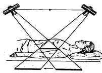
Fig. 10.14 Principle of a linear tomography
The effect of a tomography is found by continuous movement of X-ray tube and Film during exposure in opposite directions. The sharp image of an investigated layer is given only by those structures which are at the level of a rotation centre of the system "tube-Film", and structures outside of the center of this system are not visualized.
The teleroentgenography - a way of performance of roentgenography at a focal length of 150 cm and more. Due to small projective augmentation the scale of the roentgenogram approximately makes 1:1.
Polygraphy - performance of several pictures of the same organ on one Film for registration of changes of position, the form, sizes, contraction abilities of the muscular layer/3-4 a picture through 10-15-30 sec/.
Roentgenokymography - reception of the graphic representation of the contraction abilities of muscular organs by means of a special mobile lead lattice. The height of waves answers size of amplitude of reduction of a muscular organ (is shown in fig. 10.15).
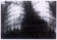
Fig. 10.15 The X-ray kymogram of a diaphragm
X-ray inspection with the use of electron optic converter of the image and videomagnetic recording - a video shooting of the x-ray image from the EOU screen.
Angiography – is a technique of a X-ray inspection of blood vessels which is peformed by entering the contrast material in to the blood vessel through a cathter after which numerous x-ray exposures are done. For exp. arteriography, venography (or a phlebography) and lymphography (see fig. 10.16). Angiography is done for the evaluation of hemodynamics, for revealing vascular pathologies, for diagnossis of the diseases caused by disturbance of the function and morphology of vessels.
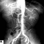

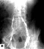
Fig. 10.16 Angiography:
a) arteriogram of the abdominal aorta and its branches; б) phlebogram of the veins of anticnemion; в) lymphogram of lymphatic vessels of ileal region
Functional roentgenography –is a method of performing roentgenograms in different functional phases of activity of organs and positions of a body
X-ray computer tomography (CТ) - the method, is based upon the measurement of a degree of weakend narrow fascicle of beams exiting from the thin layer of investigated object. The size of weakening is proportional to the size of nuclear numbers and electron density of the elements which lay on the way of narrow fascicle of the x-ray beam and depends upon its intensity and thickness of the object.
Investigation is carried out by means of a computer tomograph which consists of an X-ray tube with system of fissured collimators and detectors which contain support-gantri, a table for scanning, consoles with the equipment of regimens of the apparatus, the monitor and the computer. In the computer the signals are collected and processed which enter from the detectors: there is a digital reconstruction of the image, the information which is transferred to the console of diagnosis and management of the apparatus.
The method is based upon A.Kormakom (1963) who offered mathematical reconstruction of the level-by-level image of the brain. Haunsfield (1972) designed the first clinical model of a computer tomograph for the investigation of brain. For this scientific development in 1979 they got the Nobel Prize. In due course the computer tomograph for the investigation of the whole body of the person has been designed. Thickness of a fascicle, the layer which is allocated from the object, is possible to change depending upon the need from 1 to 10 mm.
Unlike usual roentgenography and tomography, instead of the rism detectors in the form of crystals (Natrii iodidum, etc.) or ionization gas cells (xenon) are used. Detectors perceive a difference of density of structures less than 1 % while the on x-ray Film it reaches 10-15 %. Therefore ability of detectors to perceive weakening of X-ray radiation exceeds opportunities of roentgenography by 100 times. The scheme of a x-ray computer tomograph is shown in fig. 10.17
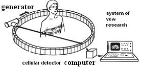
Fig. 10.17 The scheme of X-ray computer tomograph
The X-ray tube and detectors of tomographs form system which moves on a circle or on a spiral concerning investigated object. The fascicle of X-rays as a result of rotation of a tube on 180 or 360 degrees each time falls on new sites of an investigated layer and, reaching the detectors, causes an electric signal.More intensively the X-ray radiation gets on detectors, more strong electric signal they send to the computer. For identification of sites of investigated object a layer which is allocated during the tomography, are seen as the sum of identical volumes vokcel. Every vokcel has the certain projection on the matrix of computer on which numerical sizes of weakening X-ray radiations (CT-number is calculated on force of electric signals) are fixed. The plane projection of the vokcel is called as pixels, sum of these pixel forms the visual image.like roentgenogram, those sites which have appreciably weakened the X-ray radiation, will be light (bones, sites of a calcification), and those which have absorbed it a little (air,fatty tissue),However on the roentgenogram the human eye distinguishes only 16 gradation of grey color whereas in case of CТ it is possible to receive them above 1000. Size of weakening which answers density of tissues, count on scale of Haunsfields. The gradation of the scale depends upon the generation of the tomography. Density of water is seen as zero (0) size, air-1000, and bones+1000 units Haunsfield (HU). The fatty tissue has the density nearby-100 units Н, and parenchymatous organs and soft tissues - from +40 to +80 HU (see fig 10.18).
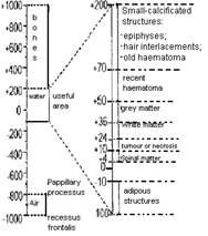
Fig. 10.18. The Haunsfilda Scale
Technique of evaluation. Appearance of a modern x-ray computer tomograph is shown in fig. 10.19.
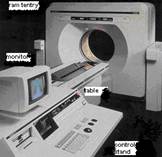
Fig. 10.19. Appearance of modern x-ray computer tomograph Tomoskan LX Philips
Support-gentri in which the X-ray tube and detectors are held has a hole in the centre. In it the table with the patient gradually linearly moves. Quantity of sections and their thickness are choosed upon there need. More thin sections give higher resolving spatial ability and accordingly allow to lead more detailed analysis and reconstruction of the image in other projections. At the same time evaluation of the certain site of a body by means of thin sections (1-2 mm) demands more time, than by means of thick (8-10 mm), that causes a greater radial load. For one section the radial load makes 0,013 Gy, and accordingly for 90 sections - 1,17 Gy. Therefore in each different case different suitable decisions are made.
In some cases to get the information of the character of pathological processes we apply intravenous contrasting which has received the name of intensifying of the image.Some pathological formations have almost same density, as well as normal tissues, they are isodense. During intravenous bolous contrasting can hold more contrast agent, than the next tissues, and look hyperdense or hypodense.
Features of the images of organs of the thorax received by roentgenography, linear tomography and computer tomography is shown in fig. 10.20.
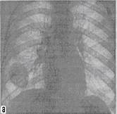
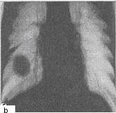
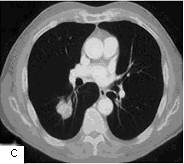
Fig 10.20 а) Survey roentgenogram of thorax in a direct projection; b) the Linear tomogram of thorax in a direct projection of the same patient with pathological formation in the right lung; c) the сomputer tomogram of thorax in an axial projection at the level of pathological formation
The x-ray spiral computer tomography - allows to receive the superfine 3 dimentional image of investigated area. Using computer tomographs with spiral scanning it is possible to receive the detailed image of appreciable anatomic site for short time and to construct its volumetric and plane reconstruction in different projections.
Дата добавления: 2015-10-23; просмотров: 140 | Нарушение авторских прав
| <== предыдущая страница | | | следующая страница ==> |
| Techniques of an X-ray investigation | | | Physical bases of MRI |