|
Читайте также: |
Figure 27. Organization of a skeletal muscle fiber

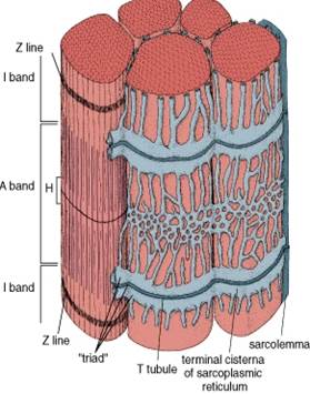
Figure 28. EM of skeletal muscle and corresponding molecular structure of sarcomere

An M line can be seen in the middle of the H band in ideal preparations.
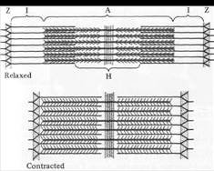
The functional unit of the myofibril is the sarcomere, the segment of the myofibril between two Z discs.
The arrangement of thick and thin filaments gives rise to the density differences that produce the cross-striations.
The thick filaments are restricted to the central portion of the sarcomere (the A band).
Thick filaments are composed of myosin. The thin filaments attach to the Z line and extend into the A band to the edge of the H zone. Portions of two sarcomeres, on either side of a Z disc, constitute the I band and contain only thin filaments.
Thick and thin filaments overlap in the lateral portions of the A band; each thick filament is surrounded by 6 thin filaments.
Contractile mechanism: sliding filament model
When a muscle contracts, each sarcomere shortens and becomes thicker, but the myofilaments remain the same length. The I band shortens during contraction, whereas the A band is unchanged in length. The H zone becomes narrower, and the thin filaments penetrate the H zone during contraction. All of these observations indicate that the thin filaments slide past the thick filaments during contraction.
Regulation of contraction: calcium, sarcoplamic reticulum and the T system
Calcium must be available for the reaction between actin and myosin, i.e., it must be available for contraction to occur. After contraction, the Ca must be removed. This rapid delivery and removal of Ca is accomplished by the combined work of the sarcoplasmic reticulum and a transverse tubular system or T system derived from the plasma membrane. The sarcoplasmic reticulum is arranged in networks around or between a group of myofilaments. One network of sarcoplasmic reticulum surrounds the A band and another network surrounds the I band. Where the two networks meet, at the junction between A and I bands the sarcoplasmic reticulum forms a slightly more regular ring like channel, called the terminal cisternae or sac, around the filaments of the myofibril.
Invaginations of the plasma membrane of the muscle cell form the T system. The T tubules penetrate to all levels of the muscle fiber. They are located between adjacent terminal cisternae.
The sarcoplasmic reticulum serves as reservoir and regulator of the Ca. Ca stimulates binding of myosin and actin. The complex of T tubule and two terminal cisternae is called the triad.
Дата добавления: 2015-07-25; просмотров: 99 | Нарушение авторских прав
| <== предыдущая страница | | | следующая страница ==> |
| Cartilage Is Capable of Two Kinds of Growth, Appositional and Interstitial | | | Contraction and its control |